Back Bones Diagram : Spinal Anatomy Center Cervical Thoracic And Lumbar Spine Info. While in the thoracic and lumbar spine, the anatomy of the vertebrae is relatively consistent between each vertebra, cervical spine anatomy is quite variable. The cervical spine consists of 7 vertebra that are numbered 1 through 7 from top to bottom i.e. The lower part of the trapezius ascends and depresses the scapula, while the transverse or middle region of the trapezius is what retracts the. Anatomical diagrams of the spine and back this human anatomy module is composed of diagrams, illustrations and 3d views of the back, cervical, thoracic and lumbar spinal areas as well as the various vertebrae. Muscle or tendon injuries can occur anywhere in the body.
The spine diagram the spine diagram shown below, consists of many bones or vertebrae,soft discs,the spinal cord, and spinal nerves. Back of skull (occipital bone) fused vertebrae (5) (sacrum) hand bones (metacarpals) finger bones (phalanges) heel bone (calcaneus) skull (cranium) backbone It connects with the collarbone at the front of the body. The vertebral column of the lower back includes the five lumbar vertebrae, the sacrum, and the coccyx. But, they are common in the back and can cause pain.
Diagram Of Vertebral Column Showing Different Parts And Regions Of The Download Scientific Diagram from www.researchgate.net The cervical spine consists of 7 vertebra that are numbered 1 through 7 from top to bottom i.e. This process continues until the end of puberty, when the growth plate stops growing and the bones fuse permanently into a single bone. The occiput (co), also known as the occipital bone, is a flat bone that forms the back of the head. Related posts of human back bones diagram human body left hand bone images. It contains the osteology, arthrology and myology of the spine and back. For more anatomy content please follow us and visit our website: The trapezius or trapezoid muscles are two paired muscles that extend from the base of the thoracic vertebrae in the spine to the occipital bone and run out to the spine of the scapula. From the front (or anterior), the vertebral body appears rounded.
Muscle or tendon injuries can occur anywhere in the body.
Human body left hand bone images 12 photos of the human body left hand bone images , bone This process continues until the end of puberty, when the growth plate stops growing and the bones fuse permanently into a single bone. The bones of the pelvis and lower back work together to support the body's weight, anchor the abdominal and hip muscles, and protect the delicate vital organs of the vertebral and abdominopelvic cavities. See lumbar spine anatomy diagram stock video clips. Bone diagram forehead (frontal bone) nose bones (nasals) cheek bone (zygoma) upper jaw (maxilla) lower jaw (mandible) breast bone (sternum) upper arm bone. These bones work together to provide. You can read more detail about these important bones in the arm from the following description and diagram. Anatomical diagrams of the spine and back this human anatomy module is composed of diagrams, illustrations and 3d views of the back, cervical, thoracic and lumbar spinal areas as well as the various vertebrae. The first seven bones (vertebrae) of your spine form your neck. The lumbar spine makes up the the lower end of the spinal column. It connects with the collarbone at the front of the body. It is designed to be incredibly strong, protecting the highly sensitive nerve roots, yet highly flexible, providing for mobility on many different planes. The low back is defined by the lumbar spine, and the pelvis is defined by the bones of the pelvic girdle.
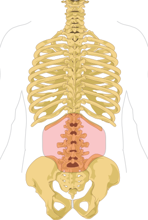
Types of scoliosis medical anatomical vector illustration diagram with spine curvatures compared with healthy back bone. These bones are connected at the back with specialized joints. Diagram of a human female skeleton, back view. The vertebral column houses the spinal canal, a cavity that. The vertebral column of the lower back includes the five lumbar vertebrae, the sacrum, and the coccyx.
Lower Back Pain Causes Herniated Disc Bulging Disc from www.afcchiropractic.com This article looks at the anatomy of the back, including bones, muscles, and nerves. 12 photos of the human back bones diagram. The vertebrae, which stack like spools of thread, support the back and protect the spinal cord. Atlas (c1) the atlas is the first cervical vertebra and therefore abbreviated c1. The bones of the pelvis and lower back work together to support the body's weight, anchor the abdominal and hip muscles, and protect the delicate vital organs of the vertebral and abdominopelvic cavities. The vertebral column of the lower back includes the five lumbar vertebrae, the sacrum, and the coccyx. Back view female with labels. Spinal anatomy is a remarkable combination of strong bones, flexible ligaments and tendons, large muscles and highly sensitive nerves.
Bone diagram forehead (frontal bone) nose bones (nasals) cheek bone (zygoma) upper jaw (maxilla) lower jaw (mandible) breast bone (sternum) upper arm bone.
Anatomynote.com found anatomy of back muscles diagram from plenty of anatomical pictures on the internet. The cervical spine consists of 7 vertebra that are numbered 1 through 7 from top to bottom i.e. The vertebral column, also known as the backbone or spine, is part of the axial skeleton.the vertebral column is the defining characteristic of a vertebrate in which the notochord (a flexible rod of uniform composition) found in all chordates has been replaced by a segmented series of bone: Vertebrae separated by intervertebral discs. Atlas (c1) the atlas is the first cervical vertebra and therefore abbreviated c1. Anatomical diagrams of the spine and back this human anatomy module is composed of diagrams, illustrations and 3d views of the back, cervical, thoracic and lumbar spinal areas as well as the various vertebrae. This vertebra supports the skull. For more anatomy content please follow us and visit our website: The bones of the pelvis and lower back work together to support the body's weight, anchor the abdominal and hip muscles, and protect the delicate vital organs of the vertebral and abdominopelvic cavities. These bones are connected at the back with specialized joints. We hope this picture anatomy of back muscles diagram can help you study and research. The lumbar spine makes up the the lower end of the spinal column. See lumbar spine anatomy diagram stock video clips.
Search for spinal column diagram. Back view female with labels. The lumbar spine connects to the thoracic spine above and the hips below. The human body is an incredible machine. This article looks at the anatomy of the back, including bones, muscles, and nerves.
Low Back Pain Wikipedia from upload.wikimedia.org The occiput (co), also known as the occipital bone, is a flat bone that forms the back of the head. Diagram of a neckbone 12 photos of the diagram of a neckbone , bone. The trapezius or trapezoid muscles are two paired muscles that extend from the base of the thoracic vertebrae in the spine to the occipital bone and run out to the spine of the scapula. C1, c2, c3, c4, etc. Tumors that begin in the bones of the spine (primary tumors) are far less common. However, the posterior bony structure is different—lamina, pedicles and bony processes project off the back of the vertebral body. It connects with the collarbone at the front of the body. Diagram of a human female skeleton, back view.
The lumbar spine connects to the thoracic spine above and the hips below.
In the back and elsewhere in the body, tendons attach muscles to bones. The red lines point individual bones and the names are writen in singular, the blue lines conect to group of bones and are in plural form. It contains the osteology, arthrology and myology of the spine and back. Human body left hand bone images 12 photos of the human body left hand bone images , bone This process continues until the end of puberty, when the growth plate stops growing and the bones fuse permanently into a single bone. The lumbar spine connects to the thoracic spine above and the hips below. The lumbar spine is composed of five vertebrae, named l1 to l5 from superior to inferior. For more anatomy content please follow us and visit our website: While in the thoracic and lumbar spine, the anatomy of the vertebrae is relatively consistent between each vertebra, cervical spine anatomy is quite variable. The vertebrae, which stack like spools of thread, support the back and protect the spinal cord. Vertebrae separated by intervertebral discs. We think this is the most useful anatomy picture that you need. However, the posterior bony structure is different—lamina, pedicles and bony processes project off the back of the vertebral body.